Neuroimaging Procedures
Neuro-Imaging Techniques NeuroCodeX™ Procedure and Process
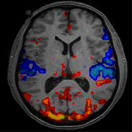
Over the years brain neuro-imaging techniques have become more refined and are being used at major institutions such as UCLA and Harvard. Brain neuro-imaging techniques fall into three major categories:
|
In an ideal world we would measure all three measurement types in our analysis, but unfortunately that can become cost prohibitive and many insurance companies will not reimburse for multiple tests.

MRI is a neuroimaging technique that is designed to look at physical/anatomical structures of the brain. It has a temporal resolution of 2 seconds-which means it is too slow to measure neuro function. Neuro function is a measure of how the brain thinks and the speed at which it processes information, which is in milliseconds.
fMRI is similar to MRI, but measures anatomical brain engagement. In other words: How well does one anatomical brain system work with another brain system? However, although it has a faster resolution, it is also too slow to measure the detailed information processing function of the brain.

CT Scan is a neuro-imaging technique and similar to MRI, but with even less resolution than MRI, which means it is too slow to measure neuro function.

PET and SPECT techniques measure metabolic activity within areas of the brain. These are gross measures, which means they are not detailed or specific measurements and cannot show neuro function.

QEEG and EEG techniques are fast enough to measure neuro function down to 100th of a millisecond, which more closely approximates brain processing speed. These faster recordings allow clearer functional measurements of brain performance with regard to thinking and processing information.
The conventional approach to brain measurements focuses on the anatomy of brain structures only, such as MRI or CAT Scans. These techniques are done to rule out basic damage to the brain structure due to severe head injuries or seizure damage. These tools and their results are not intended to be an effective measurement for mild head injuries, much less identifying neurodevelopment. These tools are too slow to measure neuro function. These methods yield great results when attempting to rule out lesions or damage to different parts of the brain’s structure. However, these tests yield little to no information concerning how well the brain is performing, especially under task or pressure.
Three other useful and popular techniques utilize fMRI, PET, or SPECT scans, which highlights anatomical, structural, or metabolic issues. fMRI is similar to MRI, but measures anatomical brain engagement. How well does one anatomical brain system work with another brain system? It has a faster resolution than MRI or CAT scan, but is still too slow to measure the detailed information processing function of the brain. PET and SPECTtechniques measure metabolic activity within areas of the brain. They are not detailed and cannot show neuro function. None of the method mentioned is fast enough to read the changes that occur in the speed of processing of the brain.
Presently, the only technique fast enough to help measure true brain processing issues is the EEG/QEEG. This method measures brain waves at the scalp’s surface. QEEG and EEGtechniques are fast enough to measure neuro function down to 100th of a millisecond, which more closely approximates brain processing speed. These faster recordings allow clearer functional measurements of brain performance with regard to thinking and processing information.
NeuroCodeX™
The NeuroCodeX™ Analysis Procedure is a proprietary process that identifies dysfunctional areas of the brain through NeuroElectic Imaging methods and modalities. The NeuroCodeX™ Report of Findings makes sense of all data collected and presents the information in a systematic, easy to understand format that helps the client understand their brain and its function. Then, based on the NeuroCodeX™ Report of Findings an appropriate integrated remediation program is recommended that can assist in correcting brain based issues.
The NeuroCodeX™ Process is firmly based on an evidence based bio-social approach to healing. We look at the individual’s neuro-function, neuro-development, physiology, and bio-chemistry and combine that with the development of the personality and various skill levels, and then finally, add in environmental effects and how each interacts with the other in order to produce the desired results.
NeuroFunction + NeuroDevelopment + Physiology + BioChemistry + Personality + Skill Level + Environment = NeuroCodeX™ Process