Brain Mapping
What is Brain Mapping?
QEEG Brain Mapping is a procedure that records electrical activity within the brain. Much like a road map, brain maps show us not only which brain systems are out of balance but also where the imbalances are located. Only by using objective measures can the brain’s processing and cognitive abilities be determined. This tool gives us the ability to view the dynamic changes taking place throughout the brain during processing tasks (such as auditory or visual processing) and assists in determining which areas of the brain are fully engaged and processing efficiently. A method of QEEG analysis called Neurometrics was developed at the Brain Research Laboratory of New York University’s Medical Center, under grant from the National Institute of Health.
Neurometric analysis aids in a more precise diagnosis of subtle brain dysfunction. It provides information about the functional organization or disorganization of the brain. It serves as a basis for identifying variations in brain function that are associated with different types of neurological disorders including Attention Deficit Disorder (ADD), Learning Disabilities (LD), Depression, Dementia, Mild Head Injury, Addiction, and Obsessive/Compulsive Disorder (OCD). This aids in determining the true underlying cause of a brain-based problem.
Neurometric analysis is FDA approved [510(k) 974748] as a diagnostic tool and presently is one of a few objective measures for many neurologically based disorders. We use this objective analysis to create more effective neuro-based protocols. This brain map provides a baseline to work from while working to overcome our client’s various issues of developmental delays, head trauma, addictions, depressions, anxiety, or learning disabilities.
Electrical Activity
 The electrical activity of the brain behaves like any other electrical system. Changes in inhibitory and excitatory synapsis potentials create voltages that are conducted through the brain. These electrical voltages enter the membranes surrounding the brain and continue up through the skull and appear at the scalp which are measured as microVolts.
These potentials are recorded by an electrode that is attached to the scalp. The electrodes are attached to an amplifier that records the electrical activity as an EEG. The EEG is recorded from many electrodes arranged in a particular pattern. This electrical activity is then compared to normative brain activity based on brain location and age.
The Stuck Brain vs Normal Functioning Brain
Symptoms such as attention and focus problems, learning and memory difficulties, and mood disorders (anxiety and depression) often occur when the brain’s processing ability is dysfunctional or damaged. Its ability to function efficiently is inadequate to do the required or desired job.
Based upon the brain’s natural ability to adapt, a damaged brain will attempt to survive by releasing neuro-inhibitors to protect it’s limited resources. In doing so it locks itself into a particular electro-chemical pattern in order to not make matters worse. When this happens, attentional flexibility may be lost, which reduces the ability to adapt to varying circumstances.
Often the personality becomes rigid and the feeling that there are no solutions to our problems. This can lead to an overwhelming sense of helplessness, hopelessness and tends to lead to mood extremes, such as depression, anger and even uncontrollable rage.
When this occurs, life in general is not fun or appealing.
What the Brain Map Numbers Mean
Brain map neurometrics and analysis allows a better view of how the brain is processing information to determine if it is performing within normal limits. Neurometrics is a mathematical technique that compares the EEGs of normal brain functioning individuals with that of the individual’s brain waves. This allows objective measures to help determine what and where the source of symptoms may be within the brain. This is especially true for those characterized by disturbances of brain organization rather than brain structures. Selected EEG features from across different brain regions quantify the organization within the brain to identify disturbances in brain processing.
Each brain map report includes extensive statistical tables of measures of absolute power and relative power, power asymmetry and synchronization (coherence), and color-coded topographic brain maps. This data is used to help identify normal and abnormal brain electrical activity.
The Specifics: How are the tables used?
When considering a set of symptoms from a brain processing point of view, the contribution of each brain wave dysfunction can often be quite revealing. Slow waves (delta waves) are generally at the root of many learning difficulties. For example, those with Dyslexia, slow brain wave activity can be found in one or more key areas. This can include the occipital lobes at the back of the brain, where incoming visual information is received and processed, Wernicke’s area located in the left Parietal lobe, where the brain processes words for understanding; Broca’s area located in the left Frontal lobe, where words are put together for expression; and often in the sensorimotor area, where speech is converted from a feeling for verbalization of thoughts.
Our brain uses its 8 13 cycles per second Alpha waves to idle itself, to rest areas not actively processing and acting on incoming sensory and motor information. While this idling is a normal and favorable phenomenon for the idling brain, if Alpha wave activity becomes “locked” and inhibited, active participation of vital brain areas cannot occur with efficiency.
The frontal lobes are the areas most commonly affected by excessive (flooding) and non-reactive(inhibited) Alpha waves. Our brain uses its frontal lobes to focus attention outside itself and to understand the complexities of the world. It is not unusual to find high amplitude, frontal Alpha in those failing academically and those having trouble meeting job demands.
To process incoming information that requires us to think and act consciously, our brain uses Beta waves. As a fast brain wave frequency it energize specific areas in the cortex. If Beta is deficient, either all over or in small areas, the brain may have insufficient energy to perform classroom or workplace tasks at peer group standards.
Keep in mind, there is no one brain wave pattern seen in all of those with a specific disease, disorder, or inefficiency. The QEEG in those with Attention Deficit Disorder, for example, may show high amplitude Delta slow waves, excessive Theta activity, or a locked in Alpha state.
Excessive Alpha and Beta brain activity is also the brain’s most reliable signature for depression, a common side effect of Learning Disabilities. Those with depression may show high Alpha or Beta, excessive coherence problems, or poor communication between the left and right frontal lobes.
Clinical symptoms of many disorders are often similar in many forms of brain processing related dysfunctions. Without the QEEG, it is almost impossible to differentiate what is what. It is the QEEG that helps the Neurotherapist sort out what is causing the symptoms in any particular individual.
Brain Wave Categories
Brain processing problems are often revealed in the QEEG in one or more of the following brain wave categories:
Absolute Power – How much Brainpower is Available?
Absolute Power measurement aids the Neurotherapist in determining whether enough brainpower within a particular frequency range is present at each recording site.
Relative Power – Who’s In Charge Here?
The Relative Power measurement aids the Neurotherapist in determining whether a particular frequency is overpowering other vital brain frequencies.
Mean Frequency – Are the Brainwaves within Spec?
Each frequency band is measured between specific ranges. The average frequency tells us whether or not that specific bandwidth is operating within normal ranges. For example the alpha frequency is measured between 8hz and 13hz. It should peak around 10 hz. Often when we observe it peaking at 9.5hz and below, we observe individuals being tired, error prone or simply misunderstanding vital input information.
Ratios – Relationships are Extremely Important!
Relationships between the various brain frequencies are compared to those individuals with normal brain functioning. Ratios lower or higher than normal are a sign of inefficiency, in either the brain’s ability to process incoming information, or of attending to and executing specific tasks.
Asymmetry – The Brain’s Balancing Act.
Asymmetry scores reveal to us whether the brain waves between the various parts of the brain are balanced. Excessive activity may indicate an overtiring of brain cells. Insufficient activity may suggest brain cells are not firing sufficiently to maintain proper brain function.
Coherence – Who’s Talking to Whom?
In order for us to understand the complexity of the world and to make and execute decisions the different parts of the brain must share information. Coherence is one of the measurements on how well the brain is able to perform this inner self-talk. This measure gives us an indication of how efficiently our brain is working to connects and disconnects different parts of itself to accomplishes a particular task.
Excessive Coherence tends to indicate two or more areas of the brain are “overly connected or locked together”. That is, the brain has become overly dependent on those centers and is not efficiently processing and executing information. This tends to result in poor day-to-day performance. Deficient Coherence means the brain is not able to efficiently connect cortical areas to perform specific tasks. Learning Disabilities may show either/or both excessive and deficient Coherence characteristics. Serious traumatic brain injury classically results in excessive Coherence.
Phase – Tortoise or the Hare?
Many of the brain’s functions are timed events, the energy from one part of the brain arriving at another area at just the right moment to perform a specific task. The QEEG measurement is called Phase. Excessive Phase statistics mean the signals arrive too early; deficient, too late. In either case, the brain is not able to do its job with peak efficiency.
The Language of the Brain
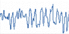
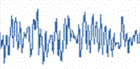
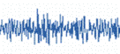
The EEG (electroencephalograph) measures brainwaves of different frequencies within the brain. Electrodes are placed on specific sites on the scalp to detect and record the electrical impulses within the brain. A frequency is the number of times a wave repeats itself within a second. It can be compared to the frequencies that you tune into on your radio. If any of these frequencies are deficient, excessive, or difficult to access, our mental performance can suffer.
- Amplitude represents the power of electrical impulses generated by brain.
- Volume or intensity of brain wave activity is measured in microvolts.
The raw EEG has usually been described in terms of frequency bands: Gamma greater than 30(Hz) BETA (13-30Hz), ALPHA (8-12 Hz), THETA (4-8 Hz), and DELTA(less than 4 Hz).
Brain Wave Frequencies
 Delta (0.1 to 3 Hz)
The lowest frequencies are delta. These are less than 4 Hz and occur in deep sleep and in some abnormal processes, also during ex-periences of “empathy state”. Delta waves are involved with our ability to integrate and let go. It reflects unconscious mind. It is the dominant rhythm in infants up to one year of age and it is present in stages 3 and 4 of sleep. It tends to be the highest in amplitude and the slowest waves. We increase Delta waves in order to decrease our awareness of the physical world. We also access information in our unconscious mind through Delta.
Peak performers decrease Delta waves when high focus and peak performance are required. However, most individuals diagnosed with Attention Deficit Disorder, naturally increase rather than decrease Delta activity when trying to focus. The inappropriate Delta response often severely restricts the ability to focus and maintain attention. It is as if the brain is locked into a perpetual drowsy state.
Another way to look at Delta is to imagine you are driving in a car and you shift into 1st gear….you’re not going to get anywhere very fast. So Delta would represent 1st gear.
|
 Theta (4-8 Hz)
The next brainwave is theta. Theta activity has a frequency of 3.5 to 7.5 Hz and is classed as “slow” activity. It is seen in connection with creativity, intuition, daydreaming, and fantasizing and is a repository for memories, emotions, sensations. Theta waves are strong during internal focus, meditation, prayer, and spiritual awareness. It reflects the state between wakefulness and sleep. These waves Relate to subconscious.
It is abnormal in awake adults but is perfectly normal in children up to 13 years old. It is also normal during sleep. Theta is believed to reflect activity from the limbic system and hippocampal regions. Theta is observed in anxiety, behavioral activation and behavioral inhibition.
When the theta rhythm appears to function normally it mediates and/or promotes adaptive, complex behaviors such as learning and memory. Under unusual emotional circumstances, such as stress or disease states, there may be an imbalance of three major transmitter systems, which results in aberrant behavior.
Back to our car example, Theta would be considered 2nd gear. Not as slow as 1st gear (Delta) but still not very fast
|
 Alpha (8-12 Hz)
Alpha waves are those between 7.5 and 13(Hz). Alpha waves will peak around 10Hz. Good healthy alpha production promotes mental resourcefulness, aids in the ability to mentally coordinate, enhances overall sense of relaxation and fatigue. In this state you can move quickly and efficiently to accomplish whatever task is at hand. When Alpha predominates most people feel at ease and calm. Alpha appears to bridge the conscious to the subconscious. It is the major rhythm seen in normal relaxed adults – it is present during most of life especially beyond the thirteenth year when it dominates the resting tracing.
Alpha rhythms are reported to be derived from the white matter of the brain. The white matter can be considered the part of the brain that connects all parts with each other. Alpha is a common state for the brain and occurs whenever a person is alert (it is a marker for alertness and sleep), but not actively processing information. They are strongest over the occipital (back of the head) cortex and also over frontal cortex.
Alpha has been linked to extroversion (introverts show less), creativity (creative subjects show alpha when listening and coming to a solution for creative problems), and mental work. When your alpha is with in normal ranges we tend to also experience good moods, see the world truthfully, and have a sense of calmness. Alpha is one of the brain’s most important frequency to learn and use information taught in the classroom and on the job. You can increase alpha by closing your eyes or deep breathing or decrease alpha by thinking or calculating.
Alpha-Theta training can create an increase in sensation, abstract thinking and self-control.
In our car scenario, Alpha would represent neutral or idle. Alpha allows us to shift easily from one task to another.
|
 Beta (above 12 Hz)
Beta activity is ‘fast’ activity. It has a frequency of 14 and greater Hz. It reflects desynchronized active brain tissue. -It is usually seen on both sides in symmetrical distribution and is most evident frontally. It may be absent or reduced in areas of cortical damage. It is generally regarded as a normal rhythm and is the dominant rhythm in those who are alert or anxious or who have their eyes open. It is the state that most of brain is in when we have our eyes open and are listening and thinking during analytical problem solving, judgment, decision making, processing information about the world around us.
Beta would represent overdrive or hyperdrive in our car scenario.
The beta band has a relatively large range, and has been divided into low, midrange and high.
Low Beta (12-15 Hz), formerly “SMR”:
|
Midrange Beta (15-18 Hz)
|
High Beta (above 18 Hz)
|
Gamma (above 36 Hz)
Gamma is measured between 36 Ð 44 (Hz) and is the only frequency group found in every part of the brain. When the brain needs to simultaneously process information from different areas, its hypothesized that the 40Hz activity consolidates the required areas for simultaneous processing. A good memory is associated with well-regulated and efficient 40Hz activity, whereas a 40Hz deficiency creates learning disabilities.
Gamma (40 Hz)
|